Mammography – Breast Imaging
At our Silverdale clinic, we provide comprehensive and individualised breast cancer screening and diagnostic services tailored to every patient’s needs. You can receive a Mammogram, Breast Ultrasound, Breast MRI, and Image-Guided Biopsy from our team of skilled and compassionate healthcare professionals.

Breast Cancer: Early detection saves lives, be proactive.
Did you know you can self-refer for a yearly screening Mammogram from the age of 40?
Know your risk
There are several scientifically validated tools available to assess an individual’s risk of breast cancer. Whilst no test is perfect for any given individual, it is a good starting point to initiate a conversation with your health practitioner who will be able to advise you at what age you should start screening and what your best screening strategy would be.
One risk calculator tool widely used in New Zealand is the: Breast Cancer Foundation NZ Risk Calculator
Know the signs and symptoms:
- Breast Lump or Thickening: A lump or thickening in the breast is one of the most common signs of breast cancer. These can be painless or tender, and they often feel different from the surrounding tissue.
- Change in Breast Size or Shape: Any unexplained change in the size or shape of your breast should be evaluated. This includes asymmetry or dimpling.
- Nipple Changes: Look out for nipple changes, such as inversion (turning inward), discharge (other than breast milk), or pain.
- Skin Changes: Watch for unusual skin changes on the breast, like redness, scaling, dimpling (resembling the skin of an orange), puckering or unexplained bruising.
- Breast Pain: While breast cancer is not typically associated with pain, persistent discomfort or pain in the breast or armpit should be investigated.
- Swelling in the Armpit or Collarbone Area: Swollen lymph nodes in these areas could be indicative of breast cancer.
If you are experiencing any of the above, don’t delay seeking your GP’s advice.
Clinical Breast Exams
Ask your healthcare provider to conduct a clinical breast exam during your regular check-ups.
Screening Mammography
It is usually recommended that women start yearly screening Mammography from the age of 40, however this can vary depending on your individual risk factors and family history. Annual Mammography should begin earlier for women at a high risk for breast cancer.
Tailored breast cancer screening recognises that every patient is unique. It is a personalised approach that ensures you receive the right screenings at the right times, enhancing your chances of early detection and effective treatment.
Discuss when to begin screenings with your healthcare provider.
For more information about breast cancer awareness, detection and support consult the Breast Cancer Foundation NZ website.
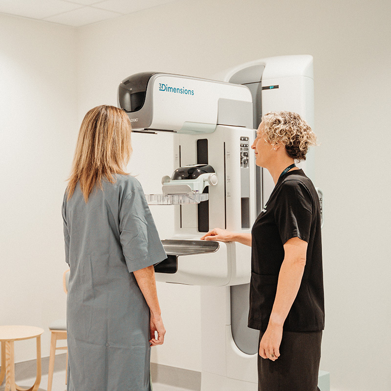
Mammography
Mammograms are the gold standard for breast cancer screening and diagnosis. At Beyond Radiology we provide the latest state of the art 3D Digital Mammography to all our patients. Our equipment is designed to detect breast abnormalities at the earliest stages, enabling us to identify potential issues even before symptoms appear.
We understand that Mammograms can be an anxiety-inducing experience. Our compassionate and caring staff is here to provide comfort and support throughout your appointment. If you have any concerns or questions, feel free to share them with us. Your well-being is our top priority.
What is 3D Mammography?
A 3D Mammogram, also known as breast tomosynthesis, is an advanced imaging tool designed to create a more detailed and comprehensive view of your breast tissue. It is an evolution of traditional 2D Mammography, which captures two images of each breast from different angles. In contrast, 3D Mammography captures a series of X-Ray images from multiple angles in 1 mm slices, which are then reconstructed to create a three-dimensional view of the breast.
What can I expect during my mammogram?
Before your appointment it is important to avoid using deodorant, talcum powder, or any creams or lotions on your breasts or underarms. These substances can interfere with the quality of the images.
We will provide a gown for you to wear during the procedure, so it is a good idea to wear a two-piece outfit for your comfort.
You will stand in front of the 3D Mammography machine and your breast will be placed on a special platform. A clear, plastic plate will gently compress your breast to ensure that the tissue is evenly spread out. While compression may cause some mild discomfort, it is essential for obtaining clear images and typically lasts only a few seconds for each view.
When can I get my results?
If you were referred by your doctor due to a new lump or any other worrisome breast symptom, your appointment will often include a Breast Ultrasound. A Specialist Breast Radiologist will conduct the ultrasound and discuss the results with you at the end of your scan. In cases where the findings appear benign, you will receive immediate reassurance. However if there are suspicious findings, a Biopsy may be recommended. Our priority is to minimize wait times and stress so we always aim to perform the Biopsy on the same day, typically guided by Ultrasound. The radiology report will be available to you and your GP on the same day. Biopsy results typically take 5-10 days to come back from the laboratory.
If you attended a routine screening Mammogram, your results are typically sent to your doctor within a few days. Sometimes we may advise for further evaluation of a specific area, which could involve additional imaging or a Biopsy. In this case your doctor will be informed, and our team will also contact you directly to arrange another appointment. You will then be seen by a Specialist Breast Radiologist who will be able to reassure you immediately or talk you through the next steps, if required.
Breast Ultrasound
This non-invasive procedure uses high-frequency sound waves to create images of breast tissue, helping us evaluate any abnormalities or concerns.
Typically, there is no special preparation required for a Breast UItrasound. You will be asked to wear a gown that can be easily adjusted to provide access to the breast.
Results will be discussed with you immediately and available to your GP on the same day if you were referred for a lump or other breast symptom. If this was a routine screening Ultrasound (in high risk patients or patients with dense breast tissue for example), the final report will be available within a few days.
Breast MRI
Why do I need a Breast MRI?
Breast cancer staging
Breast MRI is particularly beneficial for patients who have recently received a breast cancer diagnosis. It helps determine the extent of the disease, orienting the multidisciplinary team of specialists in selecting the most appropriate treatment strategy. By providing comprehensive insights into the breast tissue, it contributes to more accurate staging and treatment planning.
High-Risk Breast Cancer Screening and Abbreviated Breast MRI
Breast MRI is an essential early screening method for patients at high risk of breast cancer, for example BRCA1 or BRCA2 gene mutation carriers.
The high sensitivity of MRI is crucial for detecting cancer at its earliest stages, especially in younger patients with denser breast tissue. Dense breast tissue can reduce the sensitivity of Mammography, making MRI a valuable addition to the screening regimen.
High-risk patients, often advised by a specialist, may need to initiate their breast health screening at a younger age and undergo annual Mammograms, Ultrasounds, and Breast MRI. This comprehensive approach ensures early detection, enhancing the chances of successful treatment and improved outcomes.
High-risk patients can benefit from a shorter sequence MRI, also called abbreviated breast MRI. It is a highly sensitive screening tool to find invasive cancer at an early stage but provides less images and information than would be required for a staging MRI. The considerable advantage is that the time spent in the scanner for the patient is significantly shorter, without losing any diagnostic sensitivity. This is particularly valuable in our high risk patients who need to undergo a yearly Breast MRI.
What preparation is required for a Breast MRI scan?
For most patients, no specific preparation is required. If you are pre-menopausal and your MRI is not urgent, we may ask you to schedule your MRI between day 10-14 of your cycle.
At the time of booking and on arrival at Beyond Radiology, you will be asked to complete a safety questionnaire to ensure it is safe for you to have the scan.
Patients who have certain metal implants may be at risk of either damage to their implant or themselves. Many metal implants have been tested and are safe to enter the room however some can be dangerous such as various pacemakers.
If you have a pacemaker or metal implant within your body, it is extremely important that you tell the booking coordinator and MRI technologist at Beyond Radiology prior to your scan.
What can I expect during a Breast MRI?
You will be asked to lie face down on a comfortable padded table and your breast will be positioned within a hollow space, which is fitted with specific sensors to be able to acquire high quality images.
The examination table will slowly slide into the MRI machine, which looks like a short tunnel. You will need to remain as still as possible during the scan to obtain clear images.
In most cases, a dye (or contrast agent) will be injected through a line in your arm. This enhances the visibility of breast abnormalities and helps characterising them.
The MRI machine can produce loud knocking or thumping sounds during the scan. Ear protection is usually provided to reduce noise discomfort.
You will be in contact with a technologist throughout the procedure, as they can see and hear you through a window. You can communicate with them at any time if you have concerns or need assistance.
A Breast MRI typically takes 30-60 minutes.
The images of your scan will be available immediately after your scan to your referring doctor and the final report from the radiologist will be available within a few days. Urgent scans will be reported within a day.
If you have any questions or concerns about your upcoming Breast MRI, feel free to discuss them with your healthcare provider. Your comfort and well- being are our top priorities, and we are here to provide support and clarity throughout your breast health journey.
Breast Biopsies and Procedures
At Beyond Radiology Silverdale, we offer all range of imaged-guided breast procedures.
If an abnormality is detected on your mammogram or ultrasound, you may require a Biopsy to determine if the lesion is benign or cancerous (malignant).
All Biopsies are performed under local anaesthesia by a Specialist Breast Radiologist.
It is completely normal to feel anxious or uncertain about the procedure, so don’t hesitate to discuss any concerns with us.
Ultrasound-Guided Biopsy
Using real-time Ultrasound imaging for accurate and safe tissue sampling, this is the most frequently performed Breast Biopsy technique and is usually performed within a 30 min appointment. The Biopsy itself does not take long and is usually completed within a few minutes.
Stereotactic Biopsy
This is an advanced technique using 3D Mammography to precisely target and sample suspicious areas detected on a Mammogram but not visible on Ultrasound. The booking time is usually 45-60 min but the Biopsy itself is much shorter.
MRI-Guided Biopsy
In cases where the abnormality is only visualised with MRI, our skilled medical team can perform a targeted MRI-Guided Biopsy. This highly precise procedure involves using real-time MRI images to guide the placement of a needle for tissue sampling.
Hookwire localisation
This technique guides the surgeon in finding the area within your breast which needs to be removed during surgery when it cannot be felt on examination.
On the morning of surgery or sometimes the afternoon before your surgery, we will insert a thin flexible wire to locate this area.
The procedure will be performed using Ultrasound or X-Ray guidance, depending on how the area is best visualised.
Request an appointment
It’s easy to request your appointment with us online. Whether you’re a referrer scheduling a visit on someone’s behalf, or you’re a patient with one of our Beyond Radiology forms, simply click ‘Request an appointment’ to start the process.
Breast Cancer Risk Calculator
Estimate your 5-year risk of breast cancer now: