Intervention Radiology
Intervention Radiology is a subspecialty of radiology that uses minimally invasive image-guided procedures to diagnose and treat diseases throughout the body.

What is Intervention Radiology?
Intervention Radiology is a subspecialty of radiology that uses minimally invasive image-guided procedures to diagnose and treat diseases throughout the body.
For various musculoskeletal and sport medicine problems, subspecialist Intervention Radiologists can perform a range of intervention procedures, where the Radiologist can inject corticosteroids, Platelet Rich Plasma (PRP) or local anaesthetic, into a variety of body regions, using ultrasound, CT or fluoroscopy, in order to help alleviate symptoms and restore patient function.
Book an appointment
It’s easy to book your appointment with us online. Whether you’re a referrer scheduling a visit on someone’s behalf, or you’re a patient with one of our Beyond Radiology forms, simply click ‘Book an appointment’ to start the process.
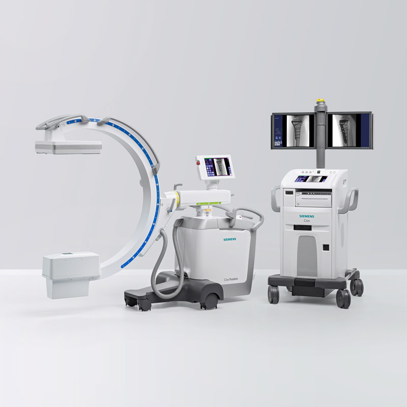
Typical CT Guided Intervention Procedures
1: Lumbar Nerve Root Injection
A nerve root injection is an image guided injection of corticosteroid and local anaesthetic around a nerve root in the spine, to decrease inflammation and help reduce pain.
What is a lumbar nerve root injection?
A subspecialist Radiologist injects a small amount of corticosteroid (steroid) and local anaesthetic into the fat surrounding a nerve root.
Corticosteroid medications decrease inflammation within and around a nerve root which may help reduce pain caused by nerve root inflammation from a variety of causes.
What is the preparation for a lumbar nerve root injection?
Limit food intake to a light meal only for up to two hours before the procedure as the procedure is performed with you lying on your front.
When booking the nerve root injection, please let the booking coordinator know if you are taking any blood thinning medication, such as aspirin, warfarin, clopidogrel and dabigatran etc.
Blood thinning medications may need to be stopped for a few days prior to the procedure, and it is very important that you do not stop or change the dose of any of these medications without consulting your doctor and the Beyond Radiology booking team.
A blood test may be required to check your blood clotting prior to the procedure.
Do not stop your normal pain medication or other medications.
You may be monitored for 1 – 2 hours after the procedure, so please allow for this.
You should arrange for a support person to drive you home after the procedure.
What happens during a lumbar nerve root injection?
You will be changed into a patient gown for comfort.
During the consent process, the Radiologist will discuss how the procedure is performed and potential risks of the procedure with you.
You will be taken into the computer tomography (CT) scanning room and asked to lie on the CT scanning table on your front. A metallic marker device will be taped to your back in the region of interest for the procedure.
The scanning table will be moved into the CT scanner and a scan performed. The Radiologist will then place a mark onto your back with a pen, where the procedure will be performed.
The Radiologist and all procedure equipment is sterile. Your back will be cleaned with a cold antiseptic, then a sterile guard placed onto your back over the cleaned area.
The Radiologist will then inject a small amount of local anaesthetic into the skin and deeper tissues of your lower back to numb this area. A sterile spinal needle will then be guided into place while taking a few CT scans to check the needle position. Once the needle is in the correct position, a small amount of contrast (dye) will be injected to check the needle position, then a combination of a water soluble corticosteroid (steroid) and long-acting local anaesthetic is injected into the tissue around the nerve root which may cause mild temporary discomfort.
How long does a lumbar nerve root injection take?
The procedure takes approximately 20 minutes.
You may be monitored after the procedure for 1 – 2 hours, so please allow for this.
Please arrange for a support person to drive you home after the procedure.
Are there any after effects of a lumbar nerve root injection?
After the procedure, you may experience mild numbness and weakness for a few hours, in your leg on the same side of your body as the injection. You will be monitored after the procedure, and not allowed to go home until the weakness in your leg has resolved.
What are the risks of a lumbar nerve root sleeve injection?
- Direct injection into the nerve root, rather than around the root, can produce intense pain and could damage the nerve, resulting in loss of sensation and weakness of the muscle supplied by the nerve.
- Injection of the medication into a surrounding blood vessel may mean the procedure does not work.
- Injection of medication into the fluid sac surrounding the spinal nerves can occur, which may cause complete numbness of both legs immediately.
- Allergic reactions to the local anaesthetic and steroid medication are rare.
- Localised infection of the skin and deep tissues is rare.
- Bleeding at the needle puncture site.
- There are very few reports in the literature of permanent leg weakness and bladder dysfunction after this procedure, which is extremely rare.
- Allergic reaction to the local anaesthetic, which may produce itching and hives or, rarely, a more severe allergic reaction.
- Allergy to the corticosteroid ranging from a mild rash to anaphylaxis is very rare, occurring in less than 1 in 250,000 patients.
- A “steroid flare” can occur which is where the person reacts to the corticosteroid, which can include redness and flushing of the face and body, usually starting a few hours or the day after the procedure and lasting a few days.
What are the benefits of a lumbar nerve root injection?
The benefits include temporary or long-term relief of back and nerve root pain which may delay or remove the need for surgery.
2: Facet Joint Injection
What is a facet joint injection?
A subspecialist Radiologist performs an injection of corticosteroid (steroid) and local anaesthetic into a facet joint of the spine, using either specialised X-Ray or computed tomography (CT) guidance, with the aim to decrease inflammation in the facet joint and reduce pain.
How do I prepare for a facet joint injection?
Limit food intake to a light meal only for up to two hours before the procedure as the procedure is performed with you lying on your front.
When booking the facet joint injection, please let the booking coordinator know if you are taking any blood thinning medication, such as aspirin, warfarin, clopidogrel and dabigatran etc.
Blood thinning medications may need to be stopped for a few days prior to the procedure, and it is very important that you do not stop or change the dose of any of these medications without consulting your doctor and the Beyond Radiology booking team.
A blood test may be required to check your blood clotting prior to the procedure.
Do not stop your normal pain medication or other medications.
You may be monitored for 1 – 2 hours after the procedure, so please allow for this.
You should arrange for a support person to drive you home after the procedure.
What happens during a facet joint injection?
You will be changed into a patient gown for comfort.
During the consent process, the Radiologist will discuss how the procedure is performed and potential risks of the procedure with you.
You will be taken into the scanning room and asked to lie on the scanning table on your front. A metallic marker device will be taped to your back in the region of interest for the procedure.
The Radiologist will then place a mark onto your back with a pen, where the procedure will be performed.
The Radiologist and all procedure equipment is sterile. Your back will be cleaned with a cold antiseptic, then a sterile guard placed onto your back over the cleaned area.
The Radiologist will then inject a small amount of local anaesthetic into the skin and deeper tissues of your lower back to numb this area. A sterile spinal needle will then be inserted under imaging guidance to check the needle position. Once the needle is in the facet joint a combination of local anaesthetic and corticosteroid will be injected into the facet joint which may produce a dull ache and mild temporary discomfort.
Are there any after effects of the facet joint injection?
The area around the facet joint is expanded during the injection which may cause mild irritation of the joint for a day or so before the steroid starts taking effect.
Rarely, there may be numbness or weakness in the arm or leg on the same side as the injection.
How long does the procedure take?
The procedure takes approximately 20 minutes.
You may be monitored after the procedure for 1–2 hours, so please allow for this.
Please arrange for a support person to drive you home after the procedure.
What are the risks of the procedure?
This is a very safe procedure.
Risks include an allergic reaction to the local anaesthetic, which may produce itching and hives or, rarely, a more severe allergic reaction.
An allergy to the corticosteroid ranging from a mild rash to anaphylaxis is very rare, occurring in less than 1 in 250,000 patients.
A “steroid flare” can occur which is where the person reacts to the corticosteroid, which can include redness and flushing of the face and body, usually starting a few hours or the day after the procedure and lasting a few days.
Some patients may experience numb arm(s) or leg(s) which may occur if the anaesthetic mixed with the steroid leaks into the adjacent epidural space. This can last up to an hour and is uncommon.
Facet joint infection is extremely rare.
Bleeding into the facet joint is very uncommon.
What are the benefits of the procedure?
After the facet joint injection, you should experience some relief of pain with approximately 80% of patients experiencing pain relief for 3 months or more.
What is the after-care following the procedure?
You may feel some minor discomfort in the area injected.
You can normally walk easily after the procedure and will be observed in the Radiology practice for approximately 20 minutes, then discharged if you are feeling well.
3: Lumbar Epidural Corticosteroid Injection
What is a lumbar epidural corticosteroid injection?
An image guided lumbar epidural corticosteroid injection is the accurate placement of a small spinal needle into the lower back region, under computed tomography (CT) guidance, so that corticosteroid (steroid) and usually a long-acting local anaesthetic can be injected into the epidural space.
What is the preparation for an epidural corticosteroid injection?
Limit food intake to a light meal only for up to two hours before the procedure as the procedure is performed with you lying on your front.
When booking the appointment, please let the booking coordinator know if you are taking any blood thinning medication, such as aspirin, warfarin, clopidogrel and dabigatran etc.
Blood thinning medications may need to be stopped for a few days prior to the procedure, and it is very important that you do not stop or change the dose of any of these medications without consulting your doctor and the Beyond Radiology booking team.
A blood test may be required to check your blood clotting prior to the procedure.
Do not stop your normal pain medication or other medications.
After the procedure, you will need to lie flat and you may be monitored by our staff for up to 2 hours, so please allow for this.
You should arrange for a support person to drive you home after the procedure.
What happens during a lumbar epidural corticosteroid injection?
You will be changed into a patient gown for comfort.
During the consent process, the Radiologist will discuss how the procedure is performed and potential risks of the procedure with you.
You will be taken into the computer tomography (CT) scanning room and asked to lie on the CT scanning table on your front. A metallic marker device will be taped to your back in the region of interest for the procedure.
The scanning table will be moved into the CT scanner and a scan performed. The Radiologist will then place a mark onto your back with a pen, where the procedure will be performed.
The Radiologist and all procedure equipment is sterile. Your back will be cleaned with a cold antiseptic, then a sterile guard placed onto your back over the cleaned area.
The Radiologist will then inject a small amount of local anaesthetic into the skin and deeper tissues of your lower back to numb this area. A sterile spinal needle will then be guided into the epidural space while taking a few CT scans to check the needle position. Once the spinal needle tip is sited in the epidural space, a combination of local anaesthetic and corticosteroid will be injected into the epidural space.
The pressure of the injection may temporarily increase the pain, which normally resolves quickly.
Are there any after-effects of a lumbar epidural corticosteroid injection?
As the injected local anaesthetic spreads throughout the epidural space, you may feel numbness in one or both legs for a short period of time.
How long does a lumbar epidural corticosteroid injection take?
The procedure takes approximately 15 to 20 minutes under CT guidance.
The time taken for full recovery is variable between 30 minutes to 2 hours.
What are the risks of a lumbar epidural corticosteroid injection?
The risks of this procedure are minimal and rare and can include:
- Increased back or leg pain during the injection, which is temporary and relatively common.
- Allergic reaction to the local anaesthetic used in the skin or mixed with the corticosteroid is very rare.
- If the thecal sac is punctured by the spinal needle which occurs in less than 5%, you may experience a severe headache for a few days.
- Bleeding into the epidural space is rare.
What are the benefits of a lumbar epidural corticosteroid injection?
The main benefits are the relief of back pain and avoidance or delay of surgery.
Typical Ultrasound Guided Intervention Procedures
1: Ultrasound Guided Bursal Injection
Is an injection of corticosteroid and local anaesthetic into a bursa, which is a soft tissue space between two structures such as bone, muscle, tendon and skin, in the shoulder, hip, knee or elbow, which allows the structures to slide over one another, in order to relieve the symptoms of bursitis.
What is a Bursal Injection?
The most commonly injected bursae are in the shoulder (subacromial bursa) and hip (trochanteric bursa).
Using a sterile technique and under ultrasound guidance, a subspecialist Radiologist will insert a sterile needle into the bursa, in order to inject a combination of corticosteroid (steroid) and local anaesthetic into the bursa.
How do I prepare for a bursal injection?
When booking the appointment, please let the booking coordinator know if you are taking any blood thinning medication, such as aspirin, warfarin, clopidogrel and dabigatran etc.
Blood thinning medications may need to be stopped for a few days prior to the procedure, and it is very important that you do not stop or change the dose of any of these medications without consulting your doctor and the Beyond Radiology booking team.
A blood test may be required to check your blood clotting prior to the procedure.
Do not stop your normal pain medication or other medications.
It is advised that you should arrange for a support person to drive you home after the procedure.
What happens during a bursal injection?
You will be changed into a patient gown for comfort and then taken into the ultrasound room and asked to lie or sit on the scanning bed, in a comfortable position.
During the consent process, an experienced sonographer (ultrasound technician) or Radiologist will explain the procedure and potential risks to you.
The Radiologist will use the ultrasound machine to locate the bursa and place a mark with a pen on the skin to indicate the site for injection.
The skin is then cleaned with antiseptic fluid.
Using a sterile technique and under ultrasound guidance, a fine needle is then advanced into the bursa. A small amount of corticosteroid and local anaesthetic is then injected into the bursa and the needle is removed.
Are there any after effects of a bursal injection?
There are minimal after effects following a bursal injection, which can include:
- a dull ache at the injection site for a few hours after the procedure.
- an area of numbness around the injection site for one or two hours after the procedure due to the local anaesthetic.
- mild bruising at the site of the injection which is rare.
For diabetics, the absorption of the corticosteroid can increase the blood sugar levels for a few days, hence it is recommended that the blood sugar levels are checked for several hours after the procedure.
How long does a bursal injection take?
The procedure normally takes approximately 5–10 minutes.
What are the risks of a bursal injection?
A bursal injection is a very safe procedure with few risks, which may include:
- temporary worsening of the symptoms over 1 to 3 days.
- allergy to the corticosteroid ranging from a mild rash to anaphylaxis is very rare, occurring in less than 1 in 250,000 patients.
- “steroid flare” can occur which is where the person reacts to the corticosteroid, which can include redness and flushing of the face and body, usually starting a few hours or the day after the procedure and lasting a few days.
- infection at the site of the injection which is rare.
- an allergic reaction to the local anaesthetic, which may produce itching and hives or, rarely, a more severe allergic reaction.
- small risk of damage to the soft tissues at the injection site called fatty atrophy (shrinking of the soft tissues) and skin pigmentation change (where the skin can go a brown colour), due to thinning or scarring of the skin or subcutaneous fat as a result of some of the injected corticosteroid leaking into the soft tissues.
What are the benefits of a bursal injection?
The aim of a bursal injection is to reduce inflammation and pain in or around the bursa by injecting a small dose of corticosteroid and local anaesthetic.
2: Ultrasound Guided Joint Injection
What is a joint injection?
A Radiologist injects a combination of a corticosteroid and local anaesthetic into a joint, under ultrasound or fluoroscopic guidance. The most common joints injected are the shoulder, knee or hip. The aim of a joint injection is to help reduce inflammation and provide pain relief.
How do I prepare for a joint injection?
No special preparation is required before a joint injection and you may eat and drink as normal.
When booking the appointment, please let the booking coordinator know if you are taking any blood thinning medication, such as aspirin, warfarin, clopidogrel and dabigatran etc.
Blood thinning medications may need to be stopped for a few days prior to the procedure, and it is very important that you do not stop or change the dose of any of these medications without consulting your doctor and the Beyond Radiology booking team.
A blood test may be required to check your blood clotting prior to the procedure.
Do not stop your normal pain medication or other medications.
It is advised that you should arrange a support person to drive you home after the procedure.
What happens during a joint injection?
You will be changed into a patient gown for comfort.
You will be taken into the imaging room and asked to lie on the bed or sit in a chair in a comfortable position.
During the consent process, an experienced Radiographer, Sonographer (ultrasound technician) or Radiologist will explain the procedure and potential risks to you.
Either an ultrasound or fluoroscopy machine will be used to locate the joint and a mark will be placed on the skin by the Radiologist to indicate the site for injection.
The skin is then cleaned with antiseptic fluid.
Using a sterile technique and under imaging guidance, a fine needle is then advanced into the joint. If the Radiologist is using the fluoroscopy machine, a small amount of radio-opaque contrast will be injected into the joint to confirm needle position within the joint.
A combination of corticosteroid and local anaesthetic is then injected into the joint and the needle is removed.
How long does a joint injection take?
An image guided joint injection normally takes between 15 to 30 minutes.
What are the benefits of a joint injection?
The aim of a joint injection is to reduce inflammation and pain.
Who performs the joint injection?
The joint injection is performed by a subspecialist Radiologist.
What are the risks of a joint injection?
This is a very safe procedure although risks can include:
- temporary worsening of the symptoms over 1 to 3 days.
- allergy to the corticosteroid ranging from a mild rash to anaphylaxis is very rare, occurring in less than 1 in 250,000 patients.
- “steroid flare” can occur which is where the person reacts to the corticosteroid, which can include redness and flushing of the face and body, usually starting a few hours or the day after the procedure and lasting a few days.
- infection at the site of the injection which is rare.
- an allergic reaction to the local anaesthetic, which may produce itching and hives or, rarely, a more severe allergic reaction.
- small risk of damage to the soft tissues at the injection site called fatty atrophy (shrinking of the soft tissues) and skin pigmentation change (where the skin can go a brown colour), due to thinning or scarring of the skin or subcutaneous fat as a result of some of the injected corticosteroid leaking into the soft tissues.
Are there any after effects of a joint injection?
- You may experience discomfort in the joint after the anaesthetic wears off after a few hours, which may last for 2-3 days after the injection.
- an area of numbness around the injection site for one or two hours after the procedure due to the local anaesthetic.
- mild bruising at the site of the injection which is rare.
For diabetics, the absorption of the corticosteroid can increase the blood sugar levels for a few days, hence it is recommended that the blood sugar levels are checked several hours after the procedure.